Electromyography
Electromyography or EMG is a diagnostic test that understands the physiological of muscles thereby assessing their health. Electromyography involves inserting a needle electrode through the skin into the muscle. This electorde detects electrical activity in the muscles and nerves controlling the muscles. A patient is asked to flex or contract the muscles so that the response of the muscle to the nerve stimuli is observed. An electromyograph is used to detect and measure electric potential that is generated by the contracting muscles. Other indicators to the proper functioning of the muscles and their corresponding nerves are the size, duration and frequency of electric signals received from them. EMG is often conducted along with a nerve conduction velocity test.
The EMG test is used to diagnose any possible weakness or impaired muscle strength due to neurological problems. Some discorders that can lead to abnormal readings on EMG test are cervical spondylosis, myasthenia gravis, carpal tunnel syndrome, myopathy, Brachial plexopathy, Guillain Barre syndrome, sciatic nerve dysfunction and mononueritis multiplex. EMG test aids in differentiating between a muscle and nerve disorder. The muscle may feel tender after the EMG test with localised bruising.
Myotonia Congenita
Myotonia Congenita is a neuromuscular genetic disease that involves progressive muscle stiffness and enlargement. This rare disorder is characterized by bouts of sustained muscle stiffness or Myotonia. The resultant muscle tensing can range from mild to severe. Myotonia Congenita occurs due to mutation of the CLCNI gene. This gene is critical in the functioning of the skeletal muscles. Therefore the mutation leads to bouts of muscle weakness. Abnormal muscle enlargement or hypertrophy is noticed in persons suffering Myotonia, even in children.
- Myotonia Congenita is an inherited condition.
- Myotonia Congenita is treatable.
- Is a non-progressive Myotonia disorder.
- Does not affect affected person's life span.
- Can affect body structure or growth patterns.
- Men are more affected than women.
- More common in northern Scandinavia, the ratio is 1:10,000 people.
- Muscle stiffness is more apparent in the leg muscles though it can affect face muscles and tongue.
- Cold, anxiety and fatigue are triggers for leg muscles stiffness.
Myotonia can affect muscles in any part of the body. In some it can affect the limbs. Some persons experience difficulty in swallowing or inability to relax their muscles quickly after contraction. Other symptoms include shortness of breath at the beginning of exercise.
Thomsen disease: In this form of the disorder, the symptoms manifest early in infancy. The main characteristic symptom of Thomsen disease is muscle stiffness or even muscle weakness after a bout of strenuous exercise. Even stress, fatigue or cold can act as triggers. The symptoms become noticeable in 2-3 years. Limbs and eyelid muscles are often affected. Thomsen disease is transmitted as an autosomal dominant trait.
Becker disease: This form of the disorder typically occurs later in life. The symptoms of Becker disease are noticed between 4-12 years. In Becker Myotonia, there is severe muscle stiffness which can often lead to mild muscle atrophy. Some medications such as beta blockers, diuretics and muscle relaxants can trigger Becker disease symptoms. Becker disease is inherited as an autosomal recessive trait.
An Electromyography is done to test the electrical activity within the muscles. Muscle biopsy is performed to check for absence of 2B fibers. CK blood test is done to check for elevated creatine kinase levels.
Myotonia Congenita Treatment
In the absence of a cure for Myotonia Congenita, a holistic approach encompassing lifestyle changes, physical therapy, and avoiding triggers is effective. Termed as the warm-up phenomenon, for most people regular exercise has been effective to relax the stiff muscles. Healthcare providers suggest making lifestyle modifications such as avoiding certain situations and physical therapy even before prescribing medications. Rehabilitative therapy to improve muscle function and relaxation techniques to reduce stress and mental discomfort which may worsen the disorder is also recommended.
Only when all these are insufficient, medications such as quinine and anticonvulsant drug such as phenytoin are prescribed. Children diagnosed with the disorder should have regular consultation with pediatric neurologist to understand and manage the disorder with the least disturbance during the growing years. Custom designed exercises, selecting suitable games and sports, avoiding foods that trigger lets children work around the symptoms and have a quality life.
Neurotmesis
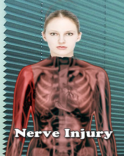
Neurotmesis etymology: Neurotmesis refers to most serious and severe nerve injury. Neurotmesis is brachial plexus injury. These brachial plexus injuries can occur in live births. The type of injury to the brachial plexus and the stretch damage will determine where the injury takes place. Various types of injuries can occur once the nerve rootlets form mixed nerve root. In some instances, the extent of the nerve damage may not be fully apparent but complete loss of motor, sensory and autonomic functions occurs. This type of complete rupture of the brachial plexus is called Neurotmesis. Neurotmesis is part of Seddon's classification scheme used to classify nerve damage. Seddon classified the nerve injury based on the extent of damage to the nerves on the basis of structural changes in cut nerves. The Seddon classification divides nerve injuries into three types namely:
Neurotmesis: Complete anatomic division of the nerve fibers with obvious discontinuity of the nerve sheath.

Axonotmesis: Microscopic division of nerve fibers without obvious discontinuity of nerve sheath.
Neuropraxia: There is injury without any anatomical discontinuity but resulting in functional disruption or nerve concussion. This is short term or sometimes lasts months with severe compression.
Neuropraxia Symptoms : Nerve Damage Symptoms: Common symptoms of Neurotmesis include loss of sensation and change in taste, expression and speech. There might be emotional and psychological disturbances. In the final stages, there could be a complete loss of motor, sensory and autonomic functions.
Diagnosis of Nerve Injury: There are many ways to diagnose the extent of the nerve injury. One of the common ways is Nerve conduction Velocity Test which tests the speed and strength of a signal being transmitted by nerve cells. Testing these factors can reveal the nature of nerve injury, such as damage to nerve cells or to the protective myelin sheath (protective coating on axons).
The test Electroneurography (EneG) which is also known as nerve conduction study or usually as a Nerve Conduction Velocity test(NCV) will help determine the nerve damage and further explore the choice of treatment.
Other than Peripheral nerve injuries, NCV is also helpful for the diagnosis of the following conditions:
Guillain Barré syndrome
Herniated disc disease
Charcot Marie Tooth disease
Special tests for assessment of Neurotmesis include electromyography, Strength duration curve, nerve conduction study and thermography. EMG test will be able to determine the presence, location and access the extent of diseases that caused the damage to the nerves and muscles. In some cases, a nerve biopsy may be needed where a small minute portion of the damaged nerve is surgically removed and analyzed.
Prognosis: Recovery from trauma is dependent on the age of the patient, type of injury and degree of injury. Without surgical intervention and repair this injury has very poor prognosis. Even with surgical repair, there could be significant loss of motor and sensory neurons which are responsible for normal conduction.
Tags: #Electromyography #Myotonia Congenita #NeurotmesisAt TargetWoman, every page you read is crafted by a team of highly qualified experts — not generated by artificial intelligence. We believe in thoughtful, human-written content backed by research, insight, and empathy. Our use of AI is limited to semantic understanding, helping us better connect ideas, organize knowledge, and enhance user experience — never to replace the human voice that defines our work. Our Natural Language Navigational engine knows that words form only the outer superficial layer. The real meaning of the words are deduced from the collection of words, their proximity to each other and the context.
Diseases, Symptoms, Tests and Treatment arranged in alphabetical order:

A B C D E F G H I J K L M N O P Q R S T U V W X Y Z
Bibliography / Reference
Collection of Pages - Last revised Date: February 25, 2026


